Blog
How to reduce the radiation dose in a CT scan? The example of the ‘Patient at Isocenter’ feature
Written by : Karen Frangie - 31/01/20 - In Blog
Even today, finding the right balance between image quality and the radiation dose remains a core issue in medical imaging. Although medical imaging is one of the most useful modern medical tools, the use of ionizing rays in imaging undeniably entails various risks.
There are many methods for reducing doses while obtaining an image quality clear enough to allow diagnosis. All of the methods for minimizing dose, optimizing dose and making adjustments enhance patient radiation protection. Dose reduction requires justification of practices and optimization of the performance of examinations. In particular, a CT scanner can reduce dose thanks to the Radiation Dose Monitor (RDM) solution.
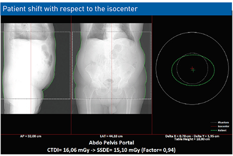
RDM’s ‘Patient at Isocenter’ feature: an important lever for reducing the dose in CT
During an examination, the CT’s technical parameters should always be adjusted to the individual patient as well as to the body region being examined. Centering the patient well is one of the strategies for optimizing the radiation dose from the scanner.
The RDM solution collects patient positioning information, enables an image to be produced accordingly, and detects whether the patient’s position is deviating from the isocenter.
Here are two points concerning RDM’s ‘Patient at Isocenter’ feature in a scanner:
– This feature assesses the patient’s centering during the examination, reinforcing good professional practices
– A shift in the patient’s position with respect to the isocenter may influence the dose
If, in the same protocol, derivations in the patient’s centering level are found, it is necessary to organize meetings with the medical staff. Training can take place with the doctors and radiographers of the institution in order to set up procedures to strengthen the level of the patient’s radioprotection.
Thanks to this feature, RDM enables the medical staff to evaluate the patient’s centering and to improve the quality of the treatment.
In Doctor Imago’s latest article “Rules and Tips to reduce the dose in CT “, Alain Noël, medical physicist, evokes the principle of the patient’s centering in the CT scan: “The automatic exposition won’t function properly if the patient is not correctly centered under the machine beforehand,” he explains. “Likewise, the mAs will have to be adapted to the patient’s body size, and modulating the dose to the organs will make it possible to deliver a lower dose to the superficial organs, such as the breast, the lens or the thyroid.”
Also, in 2018, the French public expert in nuclear and radiological risks (IRSN) published an expertise report titled: CT equipment and recommendations related to radioprotection in medical imaging. This document highlights the recommendations related to good practices, which take into account the necessities of radioprotection in medical imaging, particularly in pediatrics.
Share the post :


 Download PDF